

You can sit through a whole movie. You can drive to Oakville and back. But ten minutes on your feet at the grocery store and your legs start to ache, burn, or feel unusually heavy. You lean on the cart and it eases. You sit on a bench and it fades.
Then your MRI report arrives, filled with unfamiliar terms: lumbar spinal stenosis, central canal narrowing, foraminal stenosis at L4-L5, or nerve root compression.
So what does any of that mean for your back, your walking, and your next step?
Spinal stenosis means the spaces your nerves travel through have narrowed. Your MRI describes the structure of your spine. Your symptoms, physical examination, and everyday limitations provide the rest of the picture. Together, they help inform a personalized care plan.
Still waking up in pain after changing everything? Book a $99 spinal assessment.
What Spinal Stenosis Means on an MRI
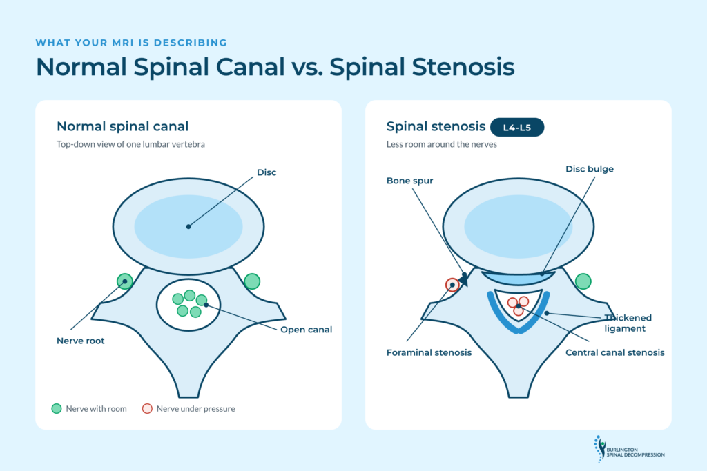
Your spinal canal is the passage through which your spinal cord and nerves travel. Smaller openings along each side, called foramina, allow individual nerves to exit the spine. Stenosis means one or more of these spaces have narrowed, sometimes putting pressure on nearby nerves.
Often, these changes develop gradually. Spinal discs lose height, joints develop arthritis, bone spurs form, and ligaments can thicken. Each change reduces the room available around the nerves.
An MRI helps show where the narrowing occurs. Common terms include:
- Central canal stenosis: Narrowing of the main spinal canal.
- Foraminal stenosis: Narrowing of an opening where a nerve root exits the spine. This can be associated with pain or tingling down one leg.
- Lateral recess stenosis: Narrowing of a passage inside the spinal canal where a nerve root travels.
- L4-L5 or L5-S1: Levels of the lower spine where the report identifies the narrowing.
The location can help explain a symptom pattern, but the scan alone cannot determine how much pain you feel or which option is best for you.
Why Walking Hurts More Than Sitting
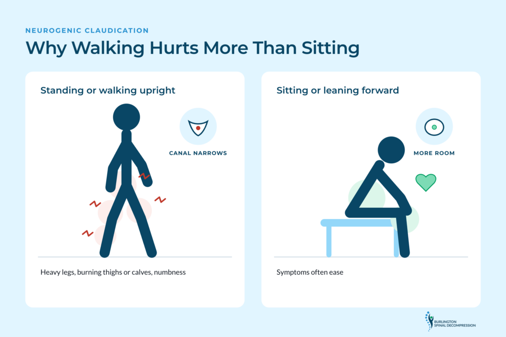
A common pattern in lumbar spinal stenosis is discomfort that gets worse with standing or walking and eases when you sit or lean forward. This is called neurogenic claudication.
When you stand upright or arch your lower back, the spaces around the nerves can become smaller. Bending forward often increases that space slightly. That helps explain why leaning on a shopping cart can feel better than walking upright.
People describe heavy legs, burning in the thighs or calves, numbness, or a feeling of unsteadiness. Symptoms can make gardening, grocery shopping, golf, or simply walking along Fairview Street more difficult.
These changes are worth discussing with the physician managing your care, especially if your walking distance is getting shorter or weakness is developing.
Why Your MRI Grade and Your Pain Do Not Always Match
Radiology reports often use terms such as mild, moderate, or severe stenosis. Those descriptions refer to the visible narrowing. They are not pain scores.
Some people with marked narrowing remain active with few symptoms. Others experience substantial limitations despite less pronounced imaging findings.
A thorough assessment therefore asks practical questions: How far can you walk? Does sitting help? Is one leg worse than the other? Have you noticed numbness, weakness, or balance changes?
These details, along with the examination and existing imaging, help connect the findings on paper to your daily life.
If your MRI mentions spinal stenosis, you can request an initial exam and bring both your report and images to discuss them in the context of your symptoms.
Non-Surgical Options for Spinal Stenosis
For many people without urgent neurological warning signs, conservative management is an appropriate place to begin. The approach depends on symptoms, examination findings, functional limitations, and medical history.
A care plan often includes several approaches working together:
- Movement and activity modification to reduce positions that aggravate symptoms.
- Guided rehabilitation to build core and hip strength and support everyday mobility.
- Walking and functional goals that can be tracked over time, such as standing longer or managing a trip to the grocery store more comfortably.
- Medical follow-up to review medicines, injections, or specialist assessment when appropriate.
The aim is to manage symptoms and preserve function, not to make promises about changing the structural findings on the MRI.
Your condition should shape more than your mattress. Understand what is happening in your spine.
Where 4D Spinal Decompression Fits
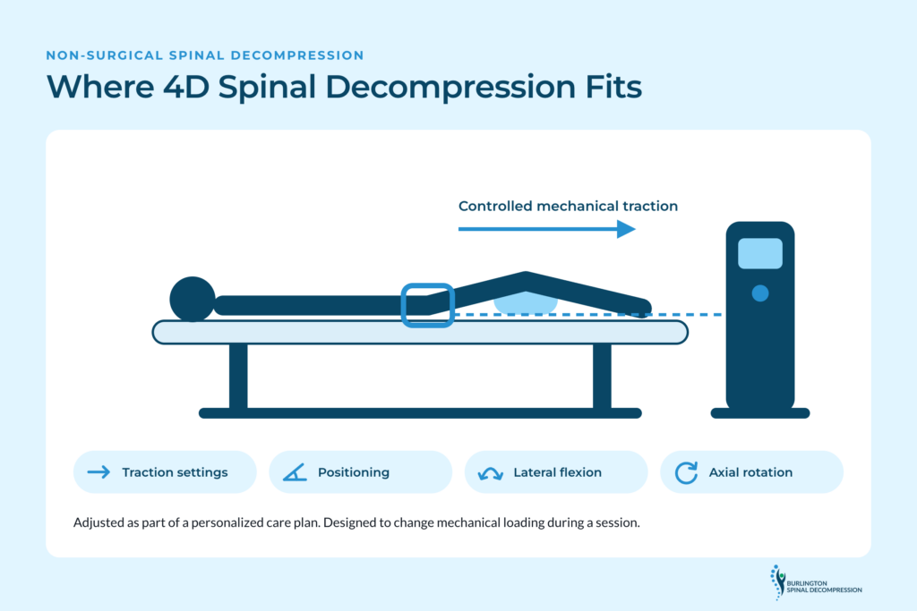
Non-surgical spinal decompression uses controlled mechanical traction. At Burlington Spinal Decompression, the clinic’s 4D spinal decompression technology allows traction settings, positioning, lateral flexion, and axial rotation to be adjusted as part of a personalized care plan.
The treatment is designed to change mechanical loading during a session. It does not remove bone spurs or reverse the underlying bony or ligament-related narrowing shown on an MRI. Evidence for traction specifically as a treatment for lumbar spinal stenosis is limited, so its potential role should be discussed in the context of an individual assessment, alongside rehabilitation and other care options.
An MRI finding at L5-S1 does not, by itself, determine a decompression program. Symptoms, examination findings, medical history, and clinical judgment also matter.
How We Approach Your MRI Results
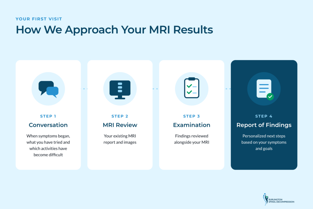
Your first visit starts with a conversation about when symptoms began, what you have tried, what makes your pain worse, and which activities have become difficult.
Dr. Brad Deakin has more than 20 years of clinical experience and understands the frustrations of persistent sciatica and lower back pain from his own experience.
The assessment includes a review of your existing MRI report and images alongside the examination findings. The radiologist’s report remains the formal interpretation of the MRI. Our assessment does not replace that interpretation or the physician overseeing your condition.
From there, the clinic discusses a Report of Findings and personalized next steps based on your symptoms, goals, and the care that is appropriate for you.
Burlington Spinal Decompression welcomes patients from Burlington, Oakville, Hamilton, Milton, Waterdown, and across Halton Region.
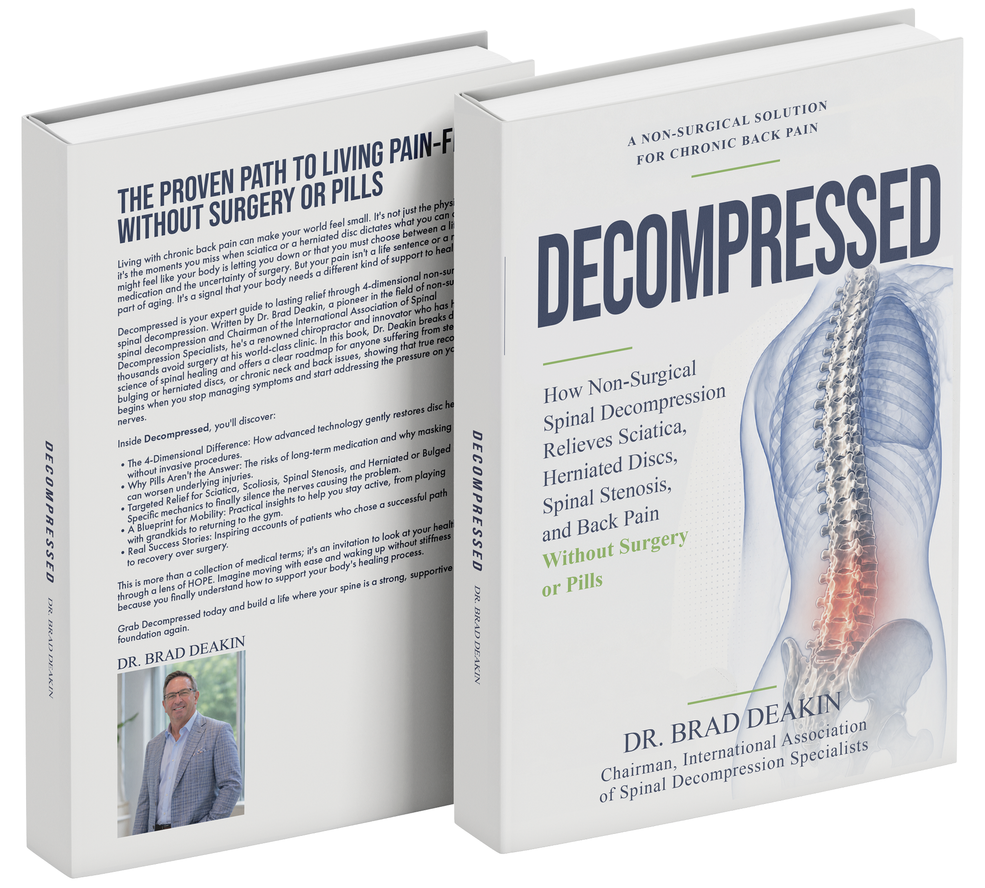
100% FREE DIGITAL COPY
Want to understand spinal decompression better?
Download a 100% free digital copy of Decompressed by Dr. Brad Deakin. The book explains sciatica, herniated discs, spinal stenosis, disc-related back pain, and how spinal decompression fits into a personalized care plan.
Get Your Free Digital CopyWhen Spinal Stenosis Needs Surgical or Urgent Medical Attention
Some people need evaluation by a spine surgeon, especially when symptoms continue to worsen despite conservative care or when significant neurological problems develop. Surgical decompression can be an appropriate option depending on the individual situation.
Seek emergency medical care immediately for new loss of bladder or bowel control, numbness around the groin or inner thighs, or sudden or rapidly worsening leg weakness. These symptoms require urgent medical evaluation, not a routine spinal decompression appointment.
Frequently Asked Questions
Bone spurs and ligament changes causing structural narrowing generally do not disappear with conservative care. However, symptoms and function can improve through suitable non-surgical management.
No. An MRI describes the structures of your spine. It does not measure pain or fully predict how stenosis affects your walking, standing, sleep, or everyday activities.
No. Although age-related changes contribute to stenosis, symptoms do not follow the same course for everyone. Some people stay stable, while others experience changes that call for a reassessment of their care.
No. An MRI describes the structures of your spine. It does not measure pain or fully predict how stenosis affects your walking, standing, sleep, or everyday activities.
Not automatically. The decision depends on your symptoms, neurological findings, limitations, overall health, and response to conservative care. Discuss the report with your physician or spine specialist.
Yes. Bring your report and, if available, the actual images on disc or through your patient imaging portal. The clinic reviews them as part of the assessment, alongside your symptoms and examination.
Your report describes what your spine looks like. Understanding what that means for standing, walking, sleeping, and enjoying your daily activities takes a wider view.
If your MRI shows spinal stenosis and you want to understand your next steps, request your initial exam with Burlington Spinal Decompression.
Or call the clinic at (289) 337-9969.
Serving Burlington, Oakville, Hamilton, Milton, Waterdown, and the Halton region.
This article provides general educational information. It does not replace medical advice, a radiologist’s imaging interpretation, or evaluation by the physician or specialist managing your care.
References
- Katz, J. N., Zimmerman, Z. E., Mass, H., & Makhni, M. C. (2022). Diagnosis and management of lumbar spinal stenosis: A review. JAMA, 327(17), 1688–1699.
- National Institute of Arthritis and Musculoskeletal and Skin Diseases: Spinal Stenosis.
- American Academy of Orthopaedic Surgeons: Lumbar Spinal Stenosis.
- Webb, C. W., Aguirre, K., & Seidenberg, P. H. (2024). Lumbar spinal stenosis: Diagnosis and management. American Family Physician, 109(4), 350–359.

